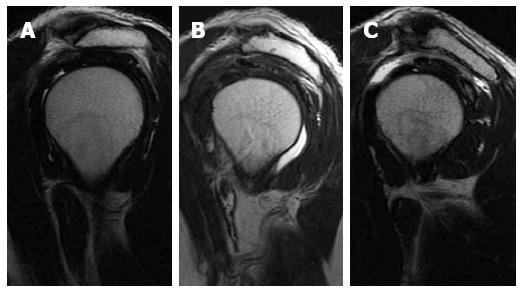
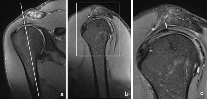
Typical magnetic resonance imaging scan showing the coracohumeral
Por um escritor misterioso
Last updated 28 maio 2024

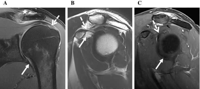
MR imaging detection of adhesive capsulitis of the shoulder: impact of intravenous contrast administration and reader's experience on diagnostic performance
Radiological anatomy: X-ray, CT, MRI
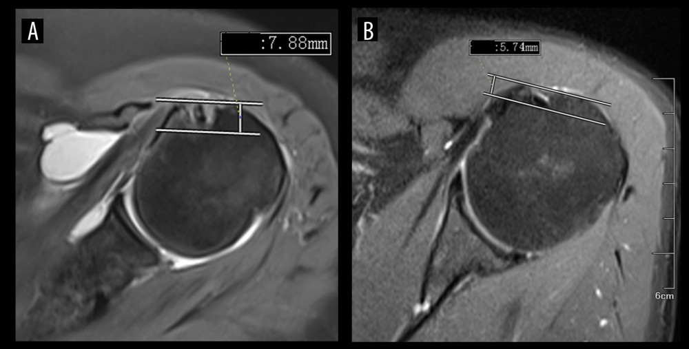
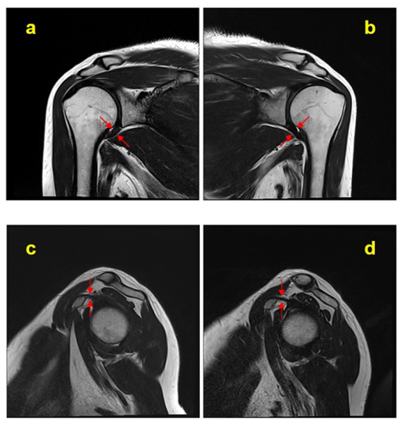
Medical Science Monitor Identification of Diagnostic Magnetic Resonance Imaging Findings in 47 Shoulders with Subcoracoid Impingement Syndrome by Comparison with 100 Normal Shoulders - Article abstract #936703
Rotator cuff disorders: How to write a surgically relevant magnetic resonance imaging report?
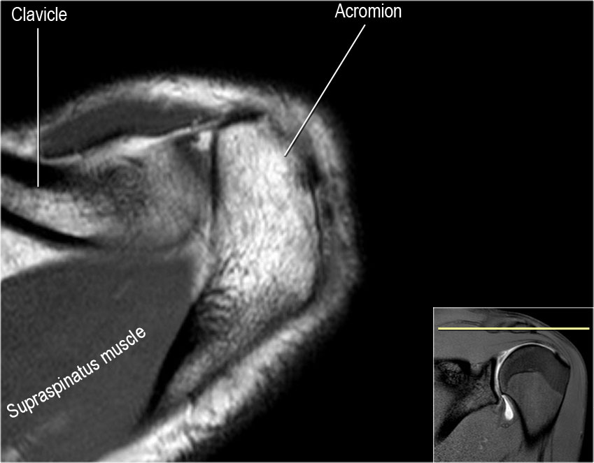
The Radiology Assistant : Shoulder Anatomy and Variants on MRI

Pain related to rotator cuff abnormalities: MRI findings without clinical significance - Bencardino - 2010 - Journal of Magnetic Resonance Imaging - Wiley Online Library
Subacromial impingement syndrome: association of multiple magnetic resonance imaging parameters with shoulder function and pain
Typical magnetic resonance imaging scan showing the coracohumeral
Contrast-enhanced Magnetic Resonance Imaging Revealing the Joint Capsule Pathology of a Refractory Frozen Shoulder
Recomendado para você
-
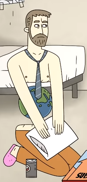
 SCP 007 : r/SCP28 maio 2024
 -
 Confinement: SCPs / Characters - TV Tropes28 maio 2024
 -
 SCP-007-J: Unidentified Muffin Creature - Site-42: SCP Foundation Fanworks (podcast)28 maio 2024
 -
 SCP-007 Abdominal Planet : r/SCP28 maio 2024
 -
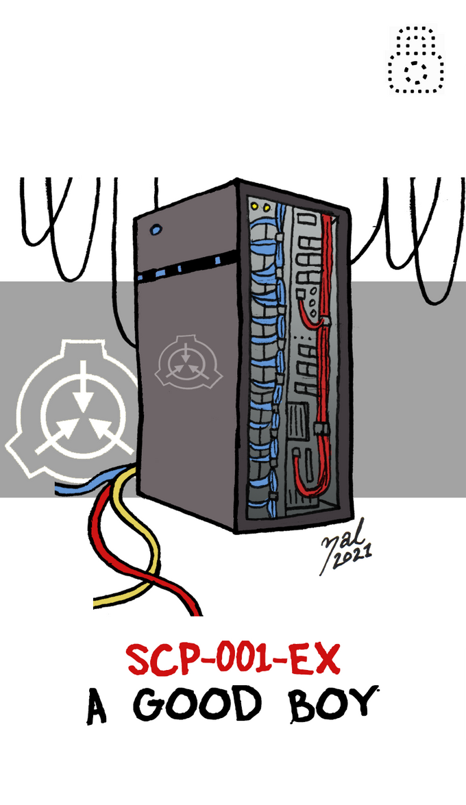
Zal Cryptid's Art Hub - SCP Foundation28 maio 2024
-
人気の「SCP-007-J」動画 2本 - ニコニコ動画28 maio 2024
-
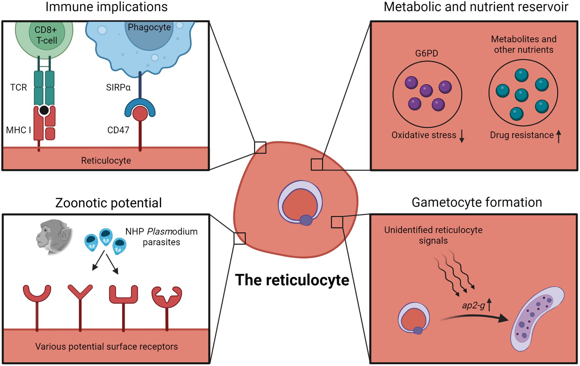 Frontiers Erythrocyte tropism of malarial parasites: The reticulocyte appeal28 maio 2024
 -
 SCP's I made Minecraft Collection28 maio 2024
 -
HLSA12,5-275 S28 maio 2024
-
scp Nova Skin28 maio 2024

você pode gostar
-
 Tata Steel Masters 2023: Ding and Abdusattorov take early lead28 maio 2024
 -
 How to Watch Auto Racing Streaming Live Today - November 1628 maio 2024
 -
 Sonic 3: Can The Film Use Shadow's Backstory?28 maio 2024
 -
 WoP in the Top 100 Indie Games 2022 – World of Padman28 maio 2024
 -
LEO Clube Sananduva28 maio 2024
-
AltBlox28 maio 2024
-
 A quick look at the Switch's new Game Boy and Game Boy Advance emulation28 maio 2024
 -
 Fantasia Sonic Infantil Com Mascara - Ri Happy28 maio 2024
 -
 Willie (Warriors), Villains Wiki28 maio 2024
 -
 novo ova do anime de Tensei Shitara Slime Datta Ken28 maio 2024

