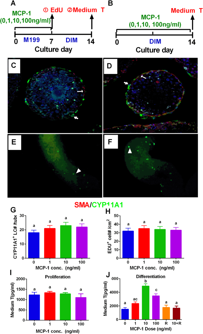
Morphology of Leydig cells in the testes after in vivo MCP-1 treatment.
Por um escritor misterioso
Last updated 17 junho 2024


Morphology of Leydig cells in the testes after in vivo MCP-1 treatment.
Monocyte Chemoattractant Protein-1 stimulates the differentiation of rat stem and progenitor Leydig cells during regeneration, BMC Developmental Biology
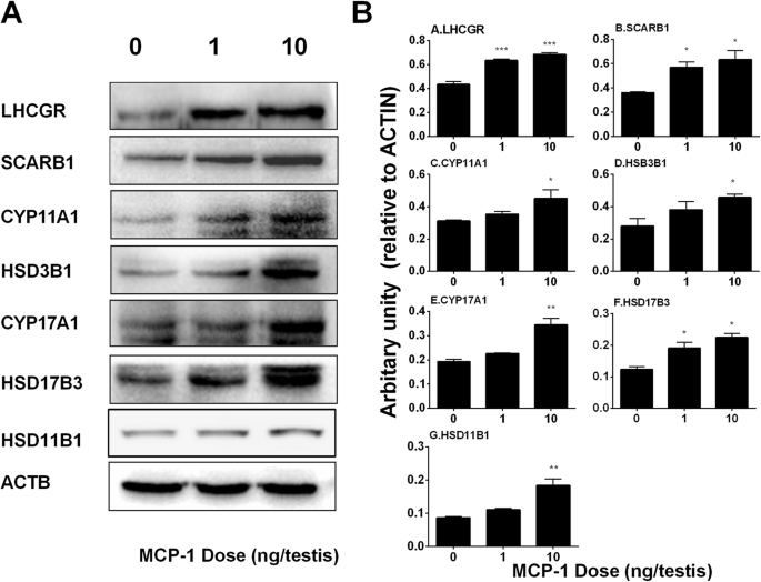
Monocyte Chemoattractant Protein-1 stimulates the differentiation of rat stem and progenitor Leydig cells during regeneration, BMC Developmental Biology

Impact of Toxoplasma gondii infection on TM3 Leydig cells: Alterations in testosterone and cytokines levels - ScienceDirect

Morphology of Leydig cells in the testes after in vivo MCP-1 treatment.

Effect of TNF on testosterone production. Leydig cells were cultured in

Transcription factor Dmrt1 triggers the SPRY1-NF-κB pathway to maintain testicular immune homeostasis and male fertility

Testicular steroidogenesis-biosynthesis of testosterone in Leydig
Frontiers Sertoli Cell Immune Regulation: A Double-Edged Sword
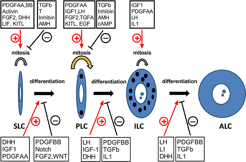
Frontiers Insights into the Development of the Adult Leydig Cell Lineage from Stem Leydig Cells
Molecules, Free Full-Text

A brief exposure to cadmium impairs Leydig cell regeneration in the adult rat testis

Testicular macrophages are recruited during a narrow time window by fetal Sertoli cells to promote organ-specific developmental functions
Recomendado para você
-
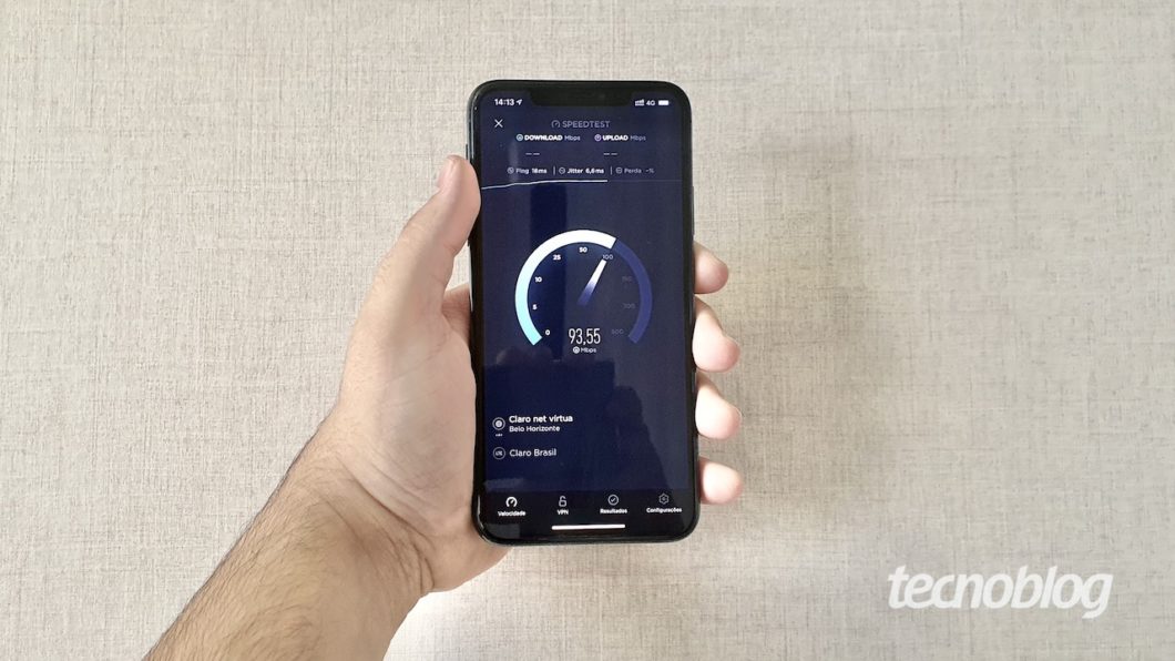
 TESTE VIVO FIBRA 300MB17 junho 2024
 -
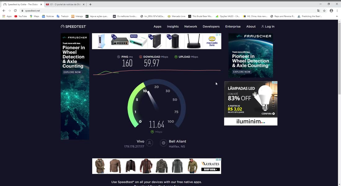
 Brasil tem 74ª internet móvel mais rápida do mundo; Claro e Vivo lideram – Tecnoblog17 junho 2024
 -
 Teste Anpad 2023 - Aula 03 de matemática financeira do curso ao vivo17 junho 2024
 -
 Teste Padrão Vivo Da Pele Do Crocodilo Do Corpo Vivo Para O Fundo Imagem de Stock - Imagem de marrom, bonito: 10245947117 junho 2024
 -
 The Division Resurgence: O próximo teste ao vivo começará em 8/1217 junho 2024
 -
 Teste Padrão Vivo Da Pele Do Crocodilo Do Corpo Vivo Para O Fundo17 junho 2024
 -
 Repórter da Record faz teste de Covid-19 ao vivo e resultado dá positivo - ISTOÉ Independente17 junho 2024
 -
 Vivo Fibra é bom? Teste do plano 300mbps/150mbps conectando a17 junho 2024
 -
 Multímetro digital Testador de caneta inteligente Caneta de capacitância de tensão de autoranging Medidor de teste elétrico Diodo-Continuidade Medidor ao vivo Testador de circuito-Sonda Ferramenta elé17 junho 2024
 -
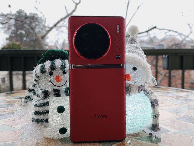 Vivo X90 Pro+ Review: Vivo sets the bar very high with its flagship smartphone - Reviews17 junho 2024
você pode gostar
-
 Call of Duty: Warzone Mobile 2.0.13545852 APK for Android17 junho 2024
 -
 10 Strongest Anime Boys Who Have White Hair17 junho 2024
 -
Manga Horse - New video is out 🥳👇17 junho 2024
-
 Butler from Zenless Zone Zero fanart : r/furry17 junho 2024
 -
/i.s3.glbimg.com/v1/AUTH_63b422c2caee4269b8b34177e8876b93/internal_photos/bs/2023/x/x/HWXz9QTgKUFjvGD23nHw/aaaaque7lfmd-kg3m6groylskvi5nddlj-ne9y3dzlxeofwx66ffsm-zf3dmnstg6tk0plbklqh-b46lk-buvsdc9pxpipsdhue9poiz65jfo40shroonkukw3xdr7wepcps8cpis54ernxs61xluw.jpg) Lançamentos da Netflix, Globoplay e Prime Video: Veja séries e filmes que estreiam em novembro, Eu17 junho 2024
Lançamentos da Netflix, Globoplay e Prime Video: Veja séries e filmes que estreiam em novembro, Eu17 junho 2024 -
Steam Workshop::UNDERTALE - Fight17 junho 2024
-
 Watcher (comics) - Wikipedia17 junho 2024
 -
arthur morgan icon|TikTok Search17 junho 2024
-
 ArmorCase-G27 Controller Case for PS5 DualSense Controller17 junho 2024
 -
 TV Academia: Rafael Leitão entrevista Giovanni Vescovi17 junho 2024


